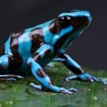
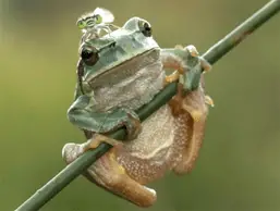
According to experts, all amphibians and reptiles found in the wild have parasites. There exists a very delicate balance of the host and the parasite and can vary greatly from the relationship of a host and parasite of creatures kept in captivity.
In nature, when an animal is not found in a small area, the concentration of parasites is low; therefore, the effects of a parasite to the host are also low. But in captive environments where there is unfavorable sanitation, the concentration of parasites can be higher and thus more dangerous.
Consider that this high concentration of parasites is affected by stress, poor husbandry, and overcrowding, any creature that is stressed or have poor immune systems will likely be affected by parasite infestation.
Before you check out the different types of parasites and how to treat them, you have to learn how parasites affect hosts and how these are detected.
Parasites may be found externally like ticks and mites or internally like digestive system worms. Parasites affect animals in all the aspects when in captivity. Generally, reptiles and amphibian pets that are affected with parasites will not live as long as other captive animals and are also more susceptible to other diseases. Also, animals that harbor parasites have a general unwell appearance. Some studies prove that animals that have a heavy parasitic infestation are unable to reproduce. And if these animals can reproduce any offspring, their babies will die at a very young age, may have stunted growth, or may have a poor growth rate.
The different parasites and ways to get rid of them
It’s very important to diagnose parasitic infections in all kinds of animals early to treat these infections properly for improved and early recovery. Vets treat parasitic infections on a case to case basis.
The different enteric parasites
Protozoa
The number of protozoa found in an amphibian is influenced by the animal’s uniqueness and the different factors that affect the gastrointestinal system. Factors like the animal’s hydration, pH, and the ability to pass the bowels affect the number and the kind of protozoa found in the animal’s digestive tract. Other factors to consider are the antagonistic relationship of different parasite species as well as predation. Protozoa can affect the type of bacteria because of predation and competition. This can have effects on the balance of the animal’s digestive system.
Flagellates
There are six genera of flagellates that live in amphibians according to experts, and these may be transmitted through cysts or by sexual intercourse. The most common flagellates that are most likely to cause problems in captive amphibians is the Hexamita. This microorganism can infect the urinary bladder and the kidneys of aquatic turtles and mostly turtles die because of severe infection. A flagellate known as Chilomastixis is found in salamanders, toads, and frogs.
Organisms like Tritrichomonas and Trichomonas are identified by their undulating membranes and are found in the intestinal tract of most reptiles and amphibians. Meanwhile, giardia is also found in amphibians, birds, and reptiles. Another common flagellum is the diplomonad flagellates found in the intestinal tract of poison dart frogs.
Ciliates
Ciliates are common in herbivorous animals. Balantidium is a common species in turtles and lizards with a herbivorous diet. But unless this organism is present in large amounts, it won’t cause any illness. But if other stresses are considered on captive animals, Balantidium can cause a severe infection. Most vets treat this ciliate according to the health condition of the animal patient.
A large ciliate called nyctotherus is present in the guts of green iguanas and turtles. The cysts of this ciliate may look similar to an egg of a trematode, and thus incorrect treatment may be provided.
Opalinids
Opalinids look similar to ciliates and flagellates, but this organism lacks a mouth and has only one nucleus. A species of opalinids called zelleriella were seen in dendrobatid frog species. And because organisms look like ciliates and flagellates, treatment may also be similar.
Amoebas
Symptoms such as anorexia, loss of weight, the presence of mucus or blood in the stool, vomiting, green color of the urates, and swellings found in the body can be due to an Entamoeba invadans infestation. This species of amoeba is very dangerous in snakes, lizards, reptiles, and amphibians. This may be diagnosed using an indirect smear of the animal’s feces.
Amoeba is commensal symbionts in animals like turtles and crocodiles. These creatures cause infection which may be transmitted to other individuals. There are also species of pathogenic amoeba, and the most common is Acanthamoeba and is responsible for most deaths. The most common symptom of acanthamoeba is star gazing which is indicative of a central nervous system condition.
Treatment of amoeba, flagellates, and ciliates
Vets have one course of action in the treatment of amoeba, flagellates, and ciliates, and this is the use of metronidazole. Vets dose up depending on the weight of the patient using a formula of 100 mg per kilogram of body weight. Doses are given by mouth and are repeated after two weeks. A dose of 50 mg per kilogram of body weight given orally daily for three to five days is also a possible course of treatment. As with all other medications given to reptiles and amphibians, every patient has to be treated uniquely. A vet uses a variety of methods to diagnose his patient before an appropriate course of treatment is provided.
Coccidia
The most common coccidian recognized in the feces of affected reptiles and amphibians is isospora and Eimeria. You can tell that an amphibian has Isospora and Eimeria when you spot listlessness, regurgitation, poor appetite, and intestinal bleeding. Aside from these, intussusception can be signs of a gall bladder or intestinal infection with coccidian.
Sarcocystis and Toxoplasma are usually found in the gut of reptiles. Affected reptiles may be the intermediate host, but reports say that snakes and lizards may be the intermediate hosts instead. Treatment for coccidia may be difficult and it is unlikely that the infection may be completely cured.
Vets use sulfamethoxine and sulfamethazine depending on the weight of the patient. The formula is usually 75 mg per kilogram of body weight given once a day for a week. Also encouraged are correct husbandry and proper hydration and sanitation for best results.
Meanwhile, cryptosporidium is most common in Biods. The typical signs are regurgitation, depression, lethargy, and swelling of the middle body. Swelling is usually seen in an advanced infestation. Cryptosporidium causes gastritis and there is still no treatment for this condition.
This microorganism is transmitted through the ingestion of sporulated oocysts as well as eating infected prey. A diagnosis may be made using a smear sample of the patient’s feces, the slime from a regurgitated food, or by getting an aspirate sample of the stomach’s contents. The latter will reveal multiple round microorganisms that are around 5 microns in diameter and will stain red. The oocysts may be passed out of the body intermittently and thus, repeated evaluations of the patient’s feces and stomach contents must be made before a definitive diagnosis is made.
If an animal is diagnosed with cryptosporidium, it has to be isolated from its other cage mates. Considerations have been made regarding cryptosporidium since treatment for this condition remains unavailable. To prevent a zoonotic threat, patients are euthanized and destroyed.
Still, some vets suggest alternative treatment like giving SMZ-TMP at 30 to 60 mg per kilogram of body weight orally for two months. Also considered is Paromomycin, given 300 to 800 mg per kilogram of weight provided orally given every 24 to 48 hours for 7 to 14 days.
Common helminths
Nematodes
Thin-walled eggs or larvae in the animal’s feces could be a sign of Stronyloides or Rhabdias infestation. The latter is found in the lungs of the hosts and will likely lead to respiratory problems. Meanwhile, Strongiloides can cause diarrhea and respiratory problems as the larvae travel to the lungs.
Both Strongyloides and Rhabdias cause poor appetite, loss of weight and inability to move properly. These creatures have a direct life cycle, and the parasites can move from host to host by ingestion of the eggs or larvae and by direct skin contact. These nematodes may also exist freely, and thus, you need to sanitize the cage of your pet frequently.
Oxyurid eggs are found in fecal exams of chelonians and lizards. Meanwhile, pinworms are non-pathogenic nematodes but are nevertheless treated by vets to avoid future problems. Oxyurids can exist in large numbers in the digestive tract, specifically in the large intestine. These microbes are present in tortoises which can cause impaction. Possible anorexia may also happen if there is a heavy infestation of Oxyurids.
Drinking contaminated water can be a way to transmit oxyurids, and this can lead to anorexia, weakness, anemia, intestinal obstruction ulceration, and peritonitis. On the other hand, capillary affects only reptiles; these settle in the intestines and also in nearby organs like the gonads and liver. The diagnosis for capillaria is through the presence of its eggs in the feces.
Ascaris is the most recognized oxyurids and are known for their strong shells. Adult Ascaris is found in the esophagus, stomach and in the small intestine where these can settle without exhibiting any sign of illness. When Ascaris are in large numbers, these can lead to regurgitation, diarrhea, and pneumonia. The most common source of Ascaris transmission is from the ingestion of intermediate hosts like rodents and amphibians.
Spirurids are another common parasite that affects the mouth and the digestive system. Reptiles are intermediate hosts or definitive hosts since terrestrial amphibians and reptiles with terrestrial amphibians, reptiles, and ants as sources of infection. Copepods are common sources in aquatic animals, and the diagnosis is through the detection of the eggs in the feces. Under the microscope, larvae are found with the egg creating a paper clip while adult worms are removed orally.
Filariae inhabits the lymph nodes, eyes, beneath the surface layers of the skin, and the colon. Microfilaria produced by adult worms may move through the blood as well as the skin, causing small tumors.
Nematode infestation treatment is done by using various anthelmintics. The most common is Piperazine at 40 to 60 mg per kilogram of weight by mouth. Other drugs are ivermectin in 0.2 mg per kilogram of body weight by mouth through an intramuscular or intradermal injection. Other medications are thiabendazole, fenbendazole, and other anti-helmentics.
Cestodes
Cestodes are tapeworms that infect reptiles and are non-host specific and hermaphroditic. Transmission of Cestodes is by ingestion of the intermediate hosts. Diagnosis of the presence of tapeworms is through the detection of eggs in the stool. Plerocercoids of Diphyllobothrium is mostly present in tadpoles that eat crustaceans.
To treat adult tapeworms, Niclosamide is given at 150 to 300 mg per kilogram of body weight orally as prescribed. Other treatment options are bunamidine HCl at 50 mg per kilogram of body weight is given orally or praziquantel at 7 to 8 mg per kilogram of weight orally or intramuscularly. For a thorough treatment, these are repeated in two weeks and are known to be effective in removing adult tapeworms.
Trematodes
Eggs found in the oral mucosa and the feces are indicative of trematode infestation. Some are found externally or found in the urinary bladder of newts, frogs, and tadpoles. The digenean trematodes create metacercariae in the skin, the eyes, and different organs. Meanwhile, the renifer group of flukes is in the mouth, esophagus, pharynx, trachea, and lungs of some snakes.
Trematodes need amphibians as intermediate hosts, therefore, taking time to prepare amphibian food or prey before giving these to your snakes. Infested animals may exhibit symptoms like anorexia or excessive salivation due to the presence of a large number of organisms in the gut and oral cavity.
To treat trematode infestation, praziquantel is given at 7 to 8 milligrams per kilogram of body weight orally, intramuscularly, and subcutaneously. Fenbendazole is also commonly used at 100 milligrams per kilogram of body weight orally. The dose is repeated after two weeks and usually, these are enough to control trematode and other metazoans.
Acanthocephalans
Acanthocephalans have a thorny head and are common in aquatic toads, turtles, and frogs. These live in the stomach and also in the intestine. Signs that your pet has acanthocephalans include the presence of blood or mucus in its stools, weight loss, and anemia.
Eggs of acanthocephalans are dark, with thick shells, and are tapered along the ends. Usually, there are fecal materials stuck to the organism’s shell. The treatment for this parasite is Levamisole at 5 to 10 mg per kilogram of body weight delivered orally, subcutaneously and ICe with doses repeated after two weeks. This is the most successful treatment.
Pentastomes
Pentastomes is a parasite that’s classified as a degenerative crustacean that affects the lungs, especially the distal air sacs in the lungs. Larvae and nymphs are deposited in the stomach wall. The transmission of pentastomes is by eating an intermediate mammalian host.
The symptoms of pentastomes are weakness, dyspnea, anorexia, and the presence of blood in the saliva. Affected animals can have this parasite in the body without exhibiting symptoms. Meanwhile, diagnosis is made through the observation of the egg that contains larva with an oval shape, with tails and four legs with pincer claws. Humans may also become hosts; therefore, you must exercise great care when handling infected animals as well as the feces of these animals. There is no known complete treatment for pentastomes but vets may prescribe levamisole at 5 mg per kilogram of weight orally or subcutaneously. Avermectins may also be given such as Milbemycin or Ivermectin.
Hemoparasites
Hemoparasites are blood parasites, and these are found in the cells or are freely floating in plasma. The development of hemoparasites involves the different body organs of their hosts. There are no clinical diseases when it comes to blood parasites but signs of possible infestation include purpura, anemia, and thrombocytopenia. Treatments for hemoparasites include tetracycline at 10 mg per kilogram of weight given orally every 24 hours and/or chloroquine phosphate at 125 mg per kilogram of weight orally every 2 days for three treatments or Quinacrine at 19 to 100 mg per kilogram of weight orally every two days for two to three weeks.
Trypanosomes
Trypanosomes are parasites that affect crocodiles, lizards, turtles, frogs, and newts. Transmission is through the bites of leeches and flies. Animals that are affected may become listless, develop a poor appetite, and may soon die due to heavy parasite levels, but pathology is often rare.
Ectoparasites
Leeches
Leeches are found in reptiles such as aquatic turtles and also in amphibians. The dorsal lymph sac and body cavity of certain species of frogs may be affected by leeches. The only way to remove leeches is by surgically removing them.
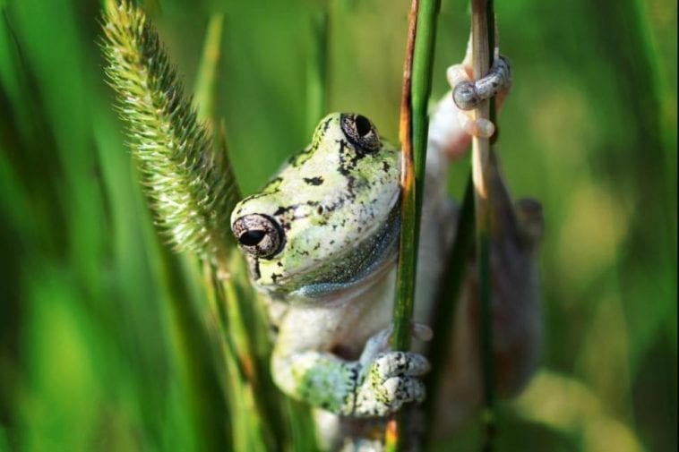
Prevention of parasites in amphibians
· Good husbandry
Cleaning and maintenance of your pet’s enclosure is a must. Your pet frog or toad likely uses a glass terrarium, and this must be cleaned at least two times a month. It should be more frequent if you have a water feature inside the terrarium.
Also included in terrarium maintenance is cleaning all supplies and accessories inside the cage. Using a good cleaner is important, or you may use natural cleaning agents like lemon, vinegar, apple cider vinegar, and baking soda.
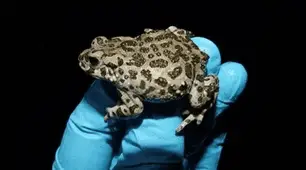
· Quarantine new animals
You should never take a new pet directly inside the cage where your other frogs are because this new guy may carry diseases and parasites. You must place it in quarantine for at least three months this is the best time to identify any possible parasites or to rule out other medical conditions.
· Regular deworming and regular vet visits
You should deworm your pets at least once a year, and if your pet is suffering from frequent infestations, then you should have it dewormed more frequently as well.
· Complete any prescribed parasite treatment
Always complete any treatment prescribed to you. If you overlook daily or weekly treatments, the parasites may still have time to multiply. Take your pet to the vet for follow up treatments right after.
Conclusion
Whether a parasite is internal or external, these can become real threats to amphibians and other animals when left undiagnosed and untreated. An effective treatment and prevention program is important. If your pet frog or toad is affected by parasites or you suspect an infestation, consult a vet right away for proper treatment.
Cleanliness and good husbandry are important in the prevention of parasites in amphibians. Individuals with weakened immune systems and those with other medical conditions must be treated right away since parasites can easily increase in number and affect these animals easily than healthy individuals. Total avoidance of parasites is impossible and difficult, especially in amphibians who eat alive food, yearly deworming and vet visits are essential.